



SWCNT as Fluorescent Probes for Multiplexed Imaging
Characterization of 17-Chirality Semiconducting Single-Walled Carbon Nanotubes via Hyperspectral Luminescence Imaging
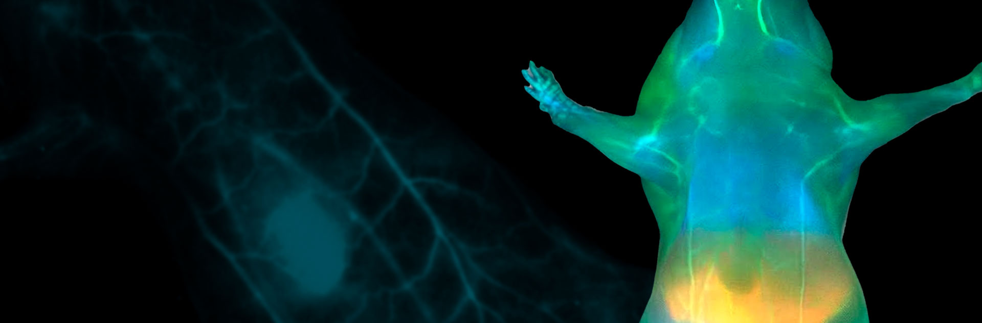
In the past few years, nondestructive optical biological imaging has grown in popularity. This approach allows visualization of cells and molecules without the needs for biopsy or cell culture. In order to obtain in-depth images of complex structures in tissue, the optical acquisition has to be performed in the second biological window (800 to 1700 nm). This spectral window provides reduced absorption, limited tissue scattering and minimal autofluorescence which greatly facilitate biological imaging.
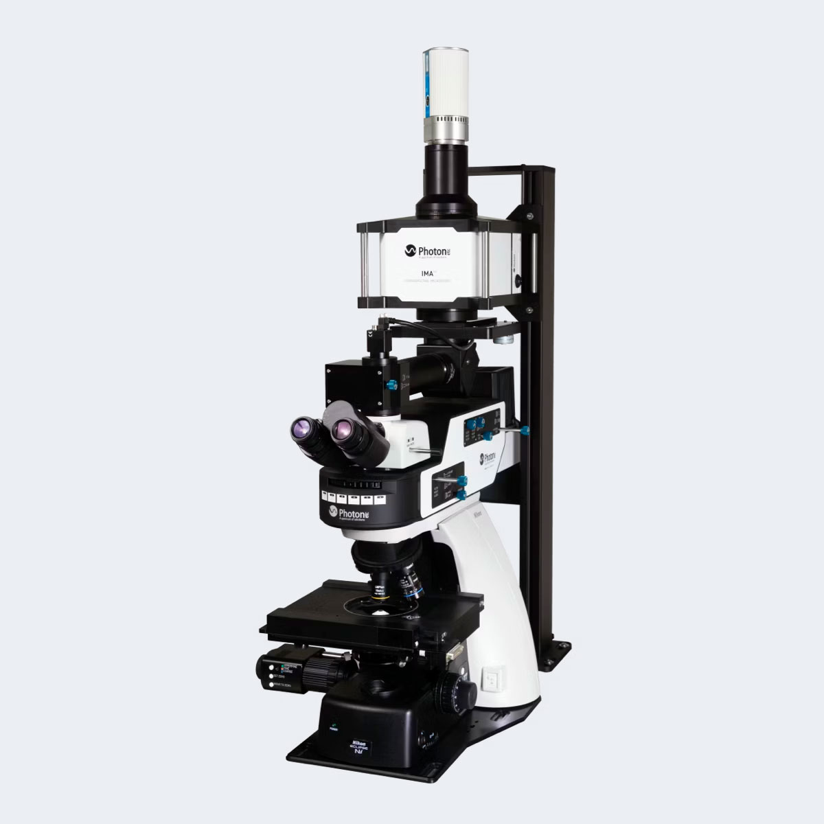
Photon etc.’s hyperspectral microscope (IMA), is an ideal tool for studies carried out in the second biological window. It is composed of a hyperspectral filter sensitive from 900 to 1620 nm coupled with a scientific microscope, a laser illumination module and an InGaAs (ZephIR or Alizé) camera. Photon etc.’s IMA platform provides spectrally and spatially resolved luminescence maps over different fields of view up to a few hundreds of micrometers square.
In order to achieve optical biological imaging, fluorescent probes are needed. Semiconducting single walled carbon nanotubes (SWCNTs) appear to be a good candidate, exhibiting excellent photostability, penetration through biological media and narrow bandwidth of emission in a wide chromatic variety. In this work from Prof. Daniel A. Heller and al. [1], SWCNTs are characterized in live cells and tissues to demonstrate their potential for multiplexed imaging applications. The fluorescence peak of emission of SWCNTs depends on each unique chiral index (n,m). In order to exploit the full potential of SWCNTs, the analytic tool used to study them needs to provide both spectral and spatial information to identify and locate those different chiralities. In this research project, 17 distinct chiralities were imaged and characterized with the help of Photon etc.’s hyperspectral microscope, IMA. Figure 1 shows an optical image of 17-chirality carbon nanotubes (a-b) with their respective luminescence spectra identified with IMA (c).
We anticipate that this approach will facilitate multiplexed nanotube imaging in biomedical applications while enabling deep-tissue optical penetration, and single-molecule resolution in vivo. [1]
Application domain
Related products
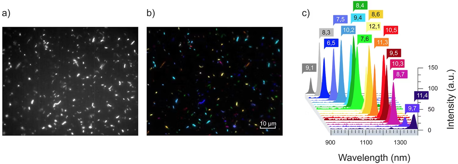
Fig. 1 – Hyperspectral microscopy of carbon nanotubes (Rice HiPco preparation) suspended with sodium deoxycholate. a) NIR broadband (900-1500 nm) fluorescence image. b) A false-color image of the same region as shown in a), colored by nanotube chirality. c) A representative spectrum of a single nanotube of each of the 17 species detected in a 500 nm emission window. Adapted from [1].
[1] Roxbury, D., Jena, P. V., Williams, R. M., Enyedi, B., Niethammer, P., Marcet, S., Verhaegen, M., Blais-Ouellette, S., & Heller, D. A. (2015). Hyperspectral Microscopy of Near-Infrared Fluorescence Enables 17-Chirality Carbon Nanotube Imaging. Scientific Reports, 5(1).
For more information, contact info@photonetc.com