



High-Contrast Diagnostic Tracking and Surgical Delineation Using Small-Molecule NIR-II Fluorophores
Standard fluorescence-guided oncological interventions frequently suffer from poor tissue penetration and intense background autofluorescence when restricted to traditional near-infrared wavelengths. To overcome these deep-tissue barriers, chemical biology has advanced toward small-molecule organic fluorophores designed specifically for the NIR-II window. This design architecture relies on extended orbital conjugation to effectively lower the energy gap, shifting emission signals into a cleaner optical spectrum where biological scattering drops precipitously.
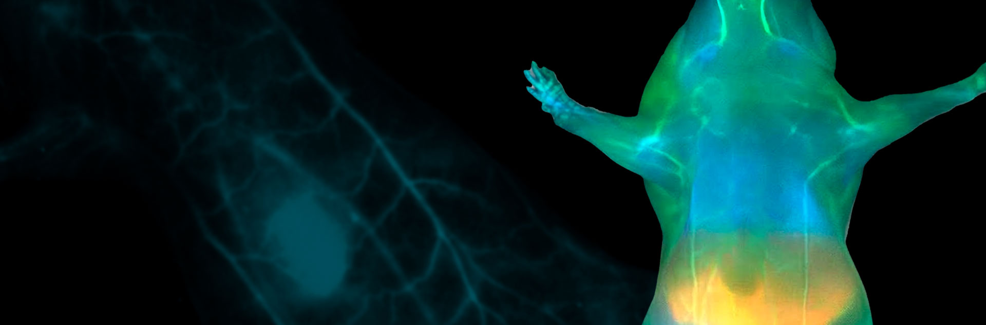
In preclinical validation models, these innovative organic tracking agents demonstrate highly localized cellular internalization combined with exceptionally favorable biocompatibility profiles. Capturing these signals requires specialized, ultra-sensitive short-wave infrared detection hardware. When paired with high-performance imaging systems, these probes yield unprecedented vascular maps and sharply defined margin outlines through multiple millimeters of scattering tissue, providing a critical roadmap to clear malignant structures while saving surrounding anatomy.
Furthermore, the adaptable and modular synthesis behind this family of dyes offers a versatile foundation for targeted therapeutics and nanobiosensor engineering. By facilitating straightforward conjugation to highly specific biological carriers or local metabolic indicators, these probes provide a stable, scalable molecular framework. This versatility bridges the gap between laboratory probe discovery and translatable imaging workflows, advancing precision oncology protocols closer to concrete diagnostic implementation.
Application domain
Related products
Supporting innovation
In advanced optical technologies
Photon etc. supports this next wave of chemical probe innovation by delivering the high-quantum-efficiency optical instruments needed to capture low-yield, long-wavelength emissions. Our specialized area-scan systems and deep-cooled NIR-II cameras allow researchers and instrumentation integrators to fully exploit the physical advantages of these novel small-molecule dyes, transforming subtle physiological signals into clear, actionable data.
To illustrate these capabilities in action, the following figure demonstrates a concrete example of real-time pharmacological mapping. The multi-panel imaging data highlights how short-wave infrared detection tracks the dynamic systemic perfusion and organ-specific accumulation of these novel small-molecule probes post-injection.

Figure adapted from npj Imaging (2026); distributed under the terms of the Creative Commons Attribution 4.0 International License (CC BY 4.0). Courtesy of the article authors.
[1] npj Imaging (Nature Portfolio), March 4, 2026. Low molecular weight 4,4′-quinocyanines for in vivo NIR-II fluorescence imaging. DOI / Source.
For more information, contact info@photonetc.com