



Clinical translation of fluorescence-guided surgery (FGS)
From Preclinical Margin Assessment to Ambient-Light Intraoperative Surgical Navigation
Precision margin assessment stands as the cornerstone of oncological resection, where completely identifying and eliminating residual malignant cells directly dictates patient survival and prevents recurrence. Traditional fluorescence-guided surgery (FGS) has historically relied on visible or first near-infrared wavelengths (NIR-I, 700–950 nm) using clinical contrast agents like indocyanine green (ICG). However, standard NIR-I methods are inherently bottlenecked by intense tissue photon scattering, limited sub-surface depth penetration, and elevated tissue autofluorescence, often obscuring small, deep-seated tumor deposits. Transitioning diagnostics into the second near-infrared window (NIR-II/SWIR, 1000–1700 nm) represents a profound paradigm shift. By operating where biological tissue scattering drops precipitously, NIR-II fluorescence unlocks sub-millimeter spatial resolution at depths several millimeters beneath the surface.
Application domain
Related products
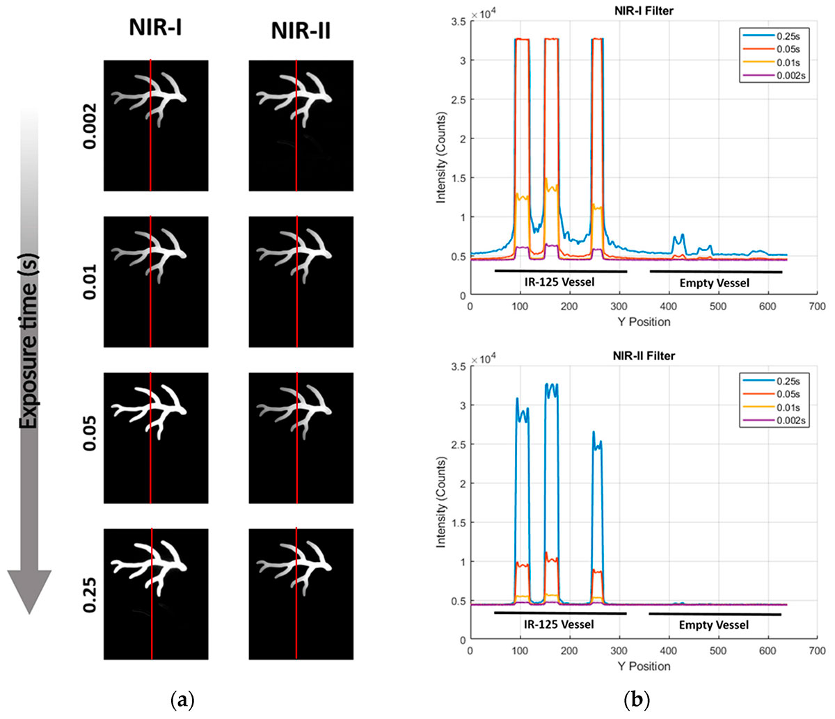
Fig. 1 – Vessel contrast for ICG in NIR-I versus NIR-II detection. (a) Representative images of a vessel phantom containing ICG-filled channels (top) and an empty channel (bottom) acquired at four exposure times (0.002, 0.01, 0.05, 0.25 s; top → bottom) with NIR-I (left) and NIR-II LP1000 (right) emission filters. Red line indicates the position of the line-profile analysis; (b) Top: NIR-I intensity profiles along the red line. Increasing exposure boosts the ICG signal (peaks) but also elevates the background and induces a false signal in the empty vessel (rightmost region). Bottom: Corresponding LP1000 profiles. ICG peaks remain well separated while baseline and the empty vessel stay at detector noise for all exposures, preserving a high SBR across the full integration range.
Translating these photophysical advantages into an active surgical workflow requires a careful balance between extreme optical sensitivity and mechanical adaptability. Back-table specimen imaging and intraoperative tracking demand systems capable of identifying targeted molecular probes or subtle ICG tail emissions with exceptional signal-to-background contrast. While highly controlled, fully dark conditions are ideal for mapping low-yield emissions, a clinical theater requires platforms that can function seamlessly under standard room lighting. Advanced hardware architectures bypass environmental light constraints by employing rapid synchronized acquisition cycles and active real-time background subtraction. This analytical framework allows surgical teams to clearly demarcate subsurface vascular networks, sentinel lymph nodes, and tumor borders without interrupting normal workflows or demanding total darkroom environments.
Dedicated infrared imaging ecosystem
Photon etc.’s dedicated infrared imaging ecosystem bridges this critical gap between lab-bench discovery and point-of-care clinical execution.
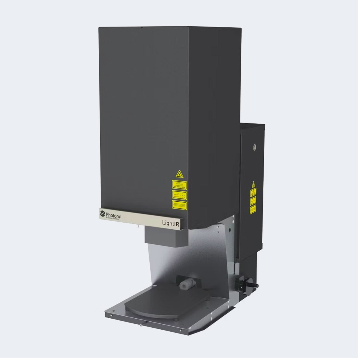
Anchored by the ultra-sensitive Alizé 1.7 InGaAs camera sensor, our solutions cater uniquely to each stage of the translational pipeline. The IR VIVO™ small-animal platform serves as the ultimate enclosed research engine, providing the high-speed, high-fidelity depth sensitivity and sub-100 µm structural resolution required to thoroughly validate novel NIR-II contrast agents and chart long-term organ biodistribution. Concurrently, the LightIR™ platform brings this exact same high-end detector technology directly into open environments. Utilizing an unshielded, portable optical head and an integrated pulse-mode configuration, the LightIR™ strips away ambient ceiling light artifacts to deliver pristine, high-contrast digital tracking deep inside open surgical fields. Together, these platforms ensure that from macro preclinical profiling to dynamic intraoperative resections, no margin is left to chance.
[1] Isuri, R. K., Williams, J., Rioux, D., Dorval, P., Chung, W., Dancer, P. A., & Delikatny, E. J. (2025). Clinical Integration of NIR-II Fluorescence Imaging for Cancer Surgery: A Translational Evaluation of Preclinical and Intraoperative Systems. Cancers, 17(16), 2676.
For more information, contact info@photonetc.com