



Advanced Intraoperative Tumor Delineation in the NIR-II Window
High-Fidelity Margin Assessment and Real-Time Preclinical Guidance via IR VIVO
Real-time oncological imaging requires optical techniques that can distinguish malignant masses from surrounding healthy structures with absolute, unambiguous clarity. Traditional fluorescence imaging in the visible or NIR-I regions often struggles with deep tissue autofluorescence and heavy photon scattering, which obscures precise tumor margins. Transitioning diagnostics into the second near-infrared window (NIR-II/SWIR, 1000–1700 nm) radically alters this landscape. By operating in a zone of minimal light-tissue interaction, NIR-II fluorescence achieves deep tissue penetration up to 10 mm alongside exceptional spatial resolution, making it a critical frontier for high-precision oncology diagnostics, lymph node mapping, and image-guided surgical resections.
The true potential of this application comes alive when paired with next-generation organic contrast platforms, such as tailored conjugated polymer nanoparticles (CPNs). Recent breakthroughs demonstrate that optimizing the backbone architecture and particle size of synthetically scalable organic polymers can unlock extraordinary optical brightness, outperforming clinical standards like indocyanine green (ICG) by several orders of magnitude. These advanced nano-emitters map out complex tumor microenvironments and tumor-associated vascular structures with pristine structural and colloidal stability. Because of their massive absorption cross-sections and superior photostability, they allow researchers to visualize micro-vasculature and define clear malignant boundaries at ultra-low minimum detectable doses, eliminating the risks of rapid signal bleaching.
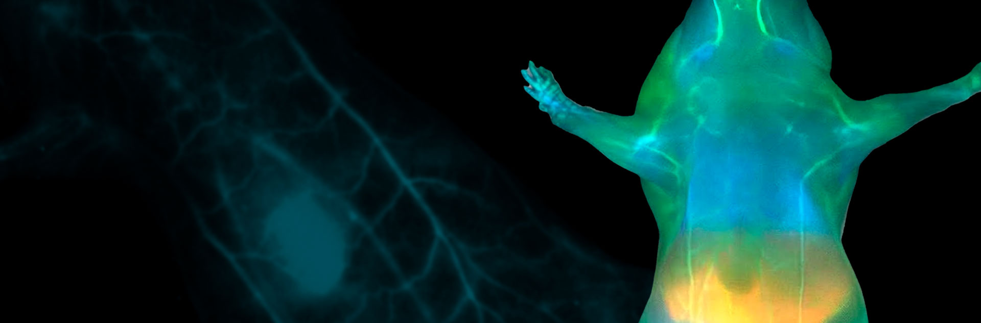
Making these profound diagnostic insights possible is Photon etc.’s IR VIVO™ whole-body preclinical imaging platform. Specifically engineered to capture weak infrared signatures with industry-leading sensitivity, the IR VIVO system utilizes ultra-low-noise detection arrays and optimized excitation-filtering combinations—such as 808 nm laser excitation paired with deep 1250 nm long-pass filtering—to isolate pure ballistic photons originating from deep within biological tissues. This enables laboratories to continuously chart real-time tumor dynamics, track organ biodistribution, and resolve fine capillary networks with remarkable signal-to-noise ratios. Whether detecting residual sub-millimeter malignant tissue during resections or evaluating the pharmacokinetics of novel theranostic agents, the IR VIVO transforms raw spectral data into high-definition, actionable physiological roadmaps.
Application domain
Related products

Fig. 1 – (A) Brightness comparison of reported NIR-I and NIR-II fluorophores, including ICG, IRDye, FD1080, Pdot-NIR1125, and PDFT-TT CPNs at two sizes (138 nm and 76 nm). (B) size stability a measured by fluctuation in the hydrodynamic diameter ($D_\text{h}$) over time (168 h) for PDFT-TT CPNs ($M_\text{w}$ = 55 kDa; $d$ = 76 nm) in buffers of varying pH. (C) Photostability of PDFT-TT CPN and ICG at the same molar concentration (0.2 nM) in phosphate-buffered saline (PBS) under continuous 808 nm excitation. In vivo fluorescence image of a mouse acquired using a 1250 nm long-pass (LP 1250) filter (D) before and (E) 60 minutes after tail vein injection of 100 μL of PDFT-TT CPNs ($M_\tex{w}$ = 55 kDa; $d$ = 76 nm). (F) Fluorescence intensity profile across a vein, acquired with an LP 1250 filter 60 minutes post-injection and Gaussian fit (blue curve). (G) Brightness comparison between ICG and PDFT-TT CPNs ($M_\text{w}$ = 55 kDa; $d$ = 76 nm) at varying concentrations, acquired with the IR VIVO animal imager using an LP 1250 filter. (H) Biodistribution of PDFT-TT CPNs ($M_\text{w}$ = 55 kDa; $d$ = 76 nm) 120 minutes after tail vein injection, showing accumulation in major organs. Images were acquired using an LP 1250 filter.
Gill, N., Posey, R., Alfaro, B., Garza, H., Tran, J., Mahmud, K. M., Fritz, A. C., Oyshi, T. A., Srivastava, I., Bickel, U., & Tropp, J. (2025). Multivariable Design of a Minimalist Conjugated Polymer Nanoparticle with Bright NIR-II Emission. ChemRxiv. DOI: 10.26434/chemrxiv-2025-h942k.
For more information, contact info@photonetc.com