



High-Resolution Angiography and Vascular Imaging in the NIR-II Spectrum
Non-Invasive Hemodynamic Profiling and High-Contrast 3D Network Reconstruction
Preclinical study of the cardiovascular network requires optical systems that can bypass the strong scattering and autofluorescence inherent to biological tissue. Photon etc.’s advanced short-wave infrared (SWIR) and NIR-II in vivo imaging platforms provide a definitive solution by operating in the 900–1700 nm optical window, where mammalian tissue achieves maximum transparency. By combining ultra-low-noise deep-cooled InGaAs camera sensors with high-intensity laser excitation, our instruments allow researchers to capture the fine architecture of blood vessels at unprecedented structural depths. This capability shifts the paradigm of optical angiography from superficial monitoring to high-contrast, sub-millimeter deep-tissue examination.
The definitive power of our platforms is highlighted when paired with state-of-the-art contrast agents, such as tailorable gold nanoclusters (AuNCs). As demonstrated in leading nanobiomedical research [1], rigidifying the surface ligands of these NIR-II emitting nanoclusters substantially shifts and optimizes their photoluminescence profile. Our high-sensitivity detection instruments seamlessly capture these enhanced emissions across specific infrared bands. Combined with deep-learning-assisted processing, this synergy enables the reconstruction of high-definition, pseudo-3D vascular networks in vivo, providing researchers with clear, high-contrast, volumetric maps of complex circulatory structures that are typically invisible to conventional NIR-I and visible setups [1].
Beyond static structural mapping, Photon etc.’s instruments deliver the rapid temporal resolution necessary to track dynamic hemodynamic events and microvascular dysfunctions in real time. For instance, in physiological models evaluating vascular compromise—such as intermittent hypoxia mimicking sleep apnea [2]—our real-time non-invasive SWIR imaging captures fast functional anomalies. It enables researchers to visualize macromolecular leakage, such as fluorescent dextran extravasation, as it passes through a disrupted blood-brain barrier or altered endothelial walls [2]. The exceptional signal-to-noise ratio provided by our instruments ensures that these subtle, time-sensitive fluorescent variations are accurately quantified from the very first seconds of injection.
Application domain
Related products

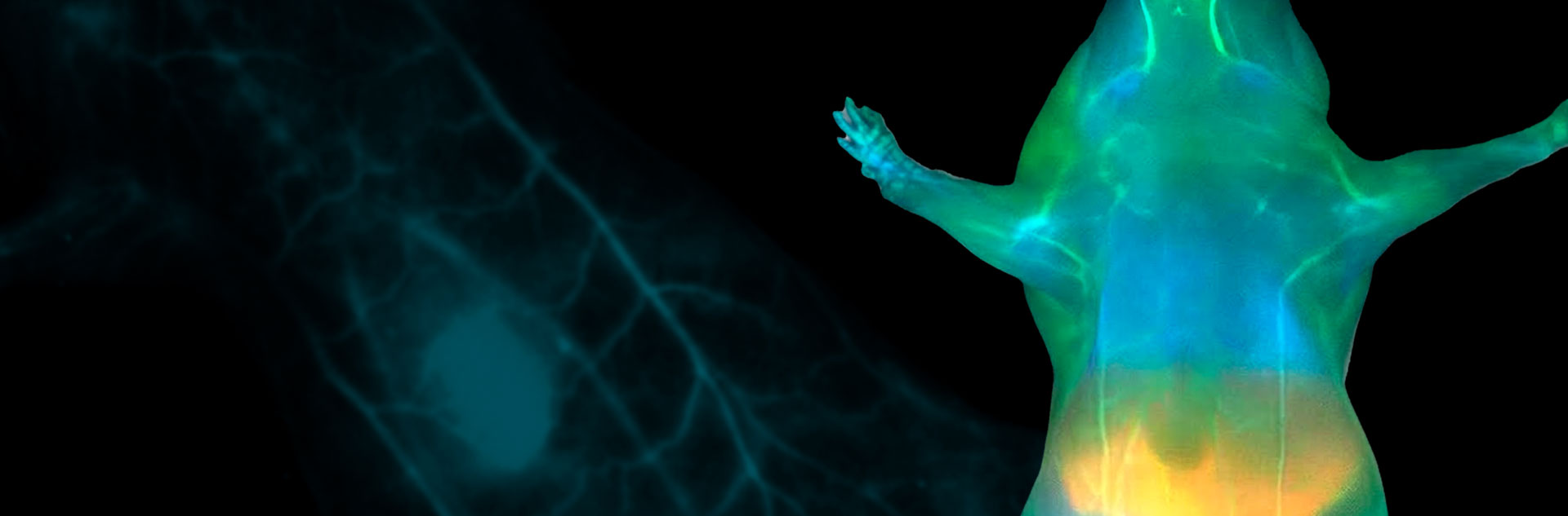
Fig. 1 – Vascular imaging in a mouse after ICG injection, acquired with a 1400 nm long-pass filter
Photon etc. provides the spectroscopic sensitivity required to advance vascular research
Engineered with flexible multi-channel emission filtering, our imaging family accommodates multiplexed fluorescent protocols, allowing laboratories to simultaneously track lymphatic drainage, macro-vascular flow, and targeted biomarker accumulation within the same animal model. Whether mapping systemic angiogenesis in oncological models, analyzing blood-brain barrier permeability, or tracking the pharmacokinetics of novel stealth theranostic nanoplatforms, Photon etc. supplies the rigorous spectroscopic sensitivity required to elevate vascular research. Our systems ensure that from microcapillary mapping to continuous perfusion tracking, every photon counts toward breakthrough physiological discovery.
[1] Le Guével, X., et al. (2022). Ligand rigidification of gold nanoclusters for enhanced NIR-II photoluminescence and deep 3D vascular reconstruction. Chemical Communications, 58, 2152-2155.
[2] Le Guével, X., et al. (2023). Real-time SWIR imaging tracks vascular dysfunction and blood-brain barrier disruption induced by intermittent hypoxia. American Journal of Physiology-Heart and Circulatory Physiology, 325(5), H1120–H1131.
For more information, contact info@photonetc.com